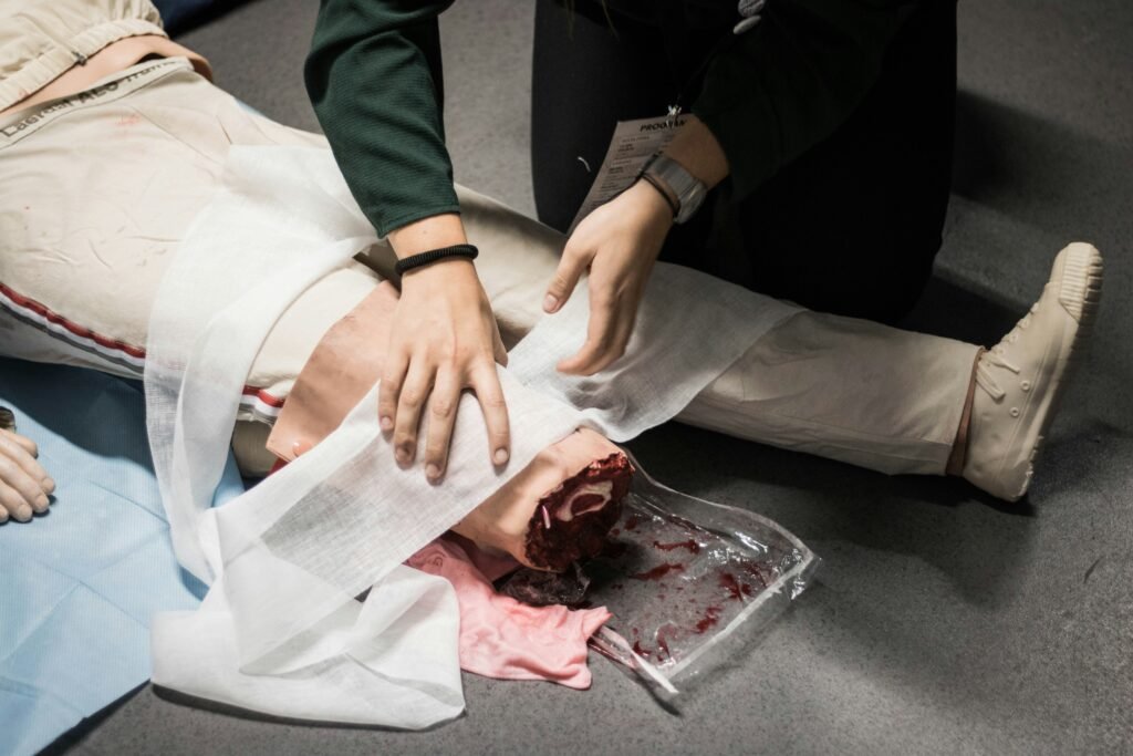
Amputation is intentional surgical removal of the limb or a body part because of some life threatening disease.
Indications of amputation
When injury or disease destroys the vitality of a part, also called a dead limb
When a local condition such as gas gangrene or extensive melanoma threatens the life of a patient.
Any trauma in which saving the life and controlling the spread is more important for example crush injuries.
Due to any infection or a sepsis.
A dying limb or ischaemic limb with late presentation
Principles
Maintain adequate blood supply to the flap.
Proper marking of the skin incision is essential
Do not use a tourniquet if performing amputation for vascular diseases.
Proximal part of the flap contains muscle component but distal part should contain only skin and deep fascia.
Flap length should be adequate; not short. It should be ideally semicircular not rectangular to get a conical stump
Pull down the nerve and cut it using a sharp knife, allowing it to retract into the soft tissue to prevent the development of neuromas.
Cut the bone with beveling and round all sharp margins.
After surgery, give active exercise to the proximal joint so that the prosthesis can fit properly to it.
Types
Non-end bearing / side bearing :The joint bears the weight.
End bearing/cone bearing : The body bears the weight.
Guillotine : Here, we do not raise any flaps; instead, we divide all tissues at the same level and keep the stump open
Formal : In this case, depending upon the indications and the decisions taken by the surgeon, and performs amputation with closure of the stump..
Different Level
Levels depends not only upon the extent of disease but also function desired in the remaining stump. This differs markedly in the upper and lower limb.
Ray : It is the amputation of toe with head of metatarsal or metacarpals.
Transmetatarsal (Gillies’) : In this case, the surgeon performs amputation proximal to the neck of the metatarsals and distal to the base.
lisfranc’s or Tarsometatarsal amputation : Here, the surgeon disarticulates the tarsometatarsal joint along with a volar flap.
But there is inevitable development of equinovarus deformity.
Stabilizing the midtarsal and ankle joints is necessary.
Chopart’s : Francis Chopart first described disarticulation through midtarsal joint. It is midtarsal amputation.
The surgeon completes the disarticulation of the foot through the talonavicular joint and the calcaneocuboid joint.
Thus, Chopart amputation removes the forefoot and midfoot, saving talus and calcaneus.
Then sutures the tibialis anterior muscle to the drilled talus bone.
Syme’s :This procedure involves excising the foot, including the calcaneum, and severing the tibia and fibula just above the ankle joint while preserving a heel flap, which necessitates dividing both malleoli.
Heel flap is supplied by medial and lateral calcaneal vessels (branches of posterior tibial artery).
Elephant boot is used for the limb after Syme’s which is inexpensive.
Pirogoff’s : It is similar to Syme’s amputation, except for the preservation of the posterior portion of the calcaneum in addition to the heel flap.
Below knee : It is also known as Burgess amputation. the ideal length of the tibial stump is 14 cm.
Peg-leg : It is amputation 5 cm below the knee level-proximal most below-knee amputation.
Hip disarticulation : When it is not possible to get minimum of l O cm length of stump of the femur, hip disarticulation is done.
This situation can occur in trauma or malignancies to get a wide clearance. Examples: Sarcomas or in cases of malignant melanomas.
Hind quarter : It is the removal of one side pelvis with innominate bone, pubis, muscles and vessels.